|
|

IMMUNOLOGY OF CANCER
Concept of Cancer Immunoediting
A variety of specific mechanisms and conditions are recruited into the tumorigenesis, including the accumulation of mutations due to physical and chemical carcinogens, epigenetic modifications like DNA methylation, metabolic disorders, inherited defects of the cell cycle and apoptosis like mutations of the TP53 gene
encoded p53, viral impacts, chronic infection, protumorigenic environment, and, as a result, breakdown of immune surveillance. Among cancer-inducing viruses, there are Epstein-Barr Virus (EBV) (Burkitt's lymphoma and Hodgkin's lymphoma), Human Herpes Virus 8 (HHV-8) (Kaposi's sarcoma), Human Papilloma Virus (HPV) (cervical cancer), Hepatitis B Virus (liver cancer), etc. In some cases, for a cancer-inducing virus, a specific cofactor is required, e.g., in Africa, malarial plasmodia contribute to the processes of development of Burkitt's lymphoma in children along with EBV. Chronic inflammation and engaged inflammatory mediators contribute to tumorigenesis by inducing gene mutations, epigenetic modifications, resistance to apoptosis in newer cancerous cells, and neoangiogenesis. Chronic inflammation appears to be associated with many cancers such as colorectal, gallbladder, hepatic, and lung cancer.
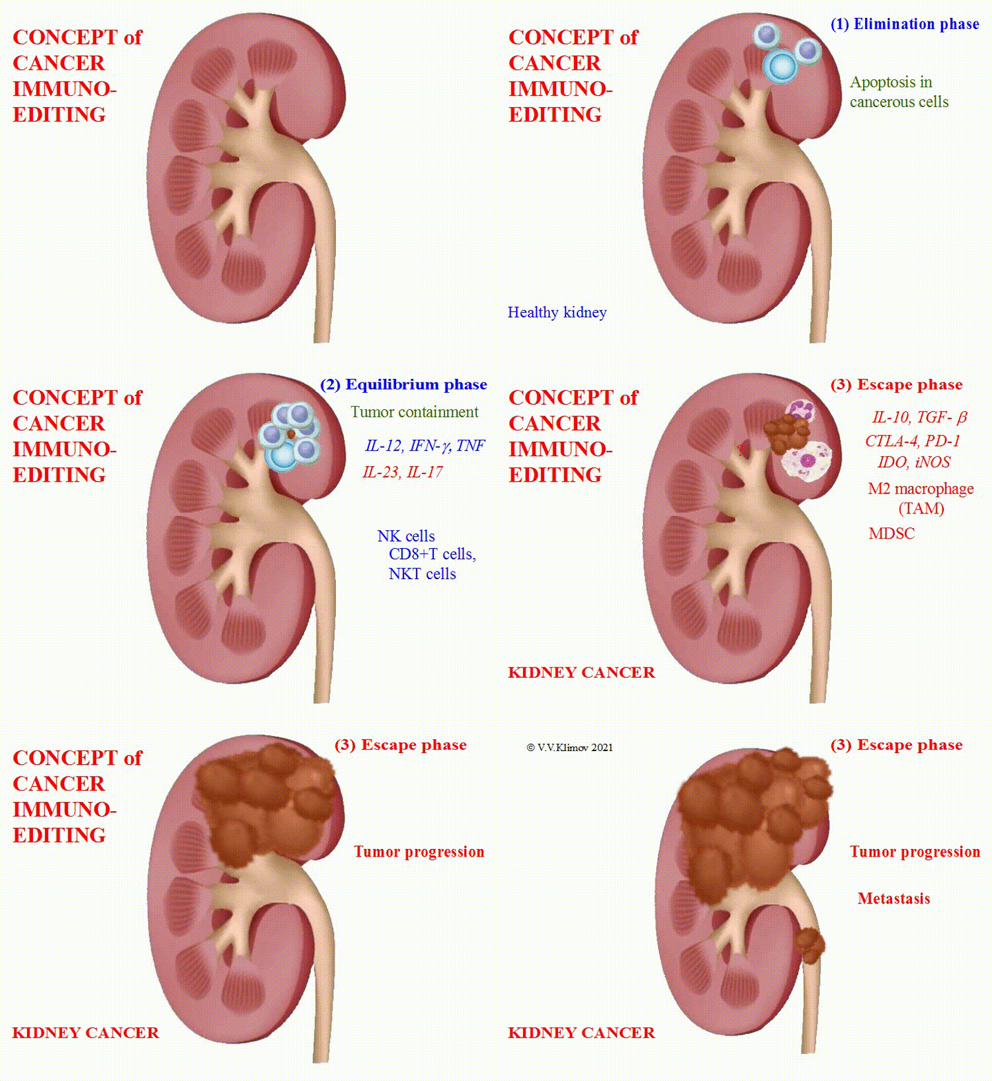
The understanding how and why the immune system misses cancer development and progression has been one of the most challenging questions in immunology. The concept of "cancer immunoediting" integrates the immune system's dual host-protective and tumor-promoting roles. Immunoediting has three major phases:
(1) elimination,
(2) equilibrium, and
(3) escape.

(1) In the course of the elimination phase, both innate immunity and adaptive immune responses are involved. Cells of the innate immune system such as NK cells, NKT cells and M1 macrophages attack a growing tumor, stimulate the production of IFN-γ, IL-12, CXCL9 (Monokine induced by IFN-γ - MIG), CXCL10 (IFN-γ-Induced Protein 10 – IP-10) and CXCL11 (IFN-Inducible T-cell α Chemoattractant – I-TAC). These chemokines promote cancerous cells' death by blocking the formation of new blood vessels. Activated NKT cells and NK cells trigger apoptosis in cancerous cells. At the same time, dendritic cells recognize tumor antigens and initiate T-cell-mediated responses. At the end of the elimination phase, CD8+T cells and CD4+T cells migrate to the tumor site, damage cancerous cells and then kill them. However, part of cancerous cells may survive.
(2) In the course of the equilibrium phase, which may last for many years, genetically unstable and rapidly mutating cancerous cells consequently, acquire partial resistance to the immune system. It isshown that the balance of IL-12 promoting cancer elimination and IL-23 promoting cancer persistence maintains tumors in equilibrium. Respectively, since IL-23 is an upregulator of IL-17 production, Th17 may play a negative role in the maintenance of equilibrium. In addition, the recognition of Tumor-Associated Molecular Patterns (TAMPs) by PRRs may lead to chronic inflammation favoring tumor progression. Also, high proportions of CD8+ T cells, NK cells, γδT cells and low ratios of NKT cells, pTreg cells, and Myeloid-Derived Suppressor Cells (MDSCs) are found during this phase. Importantly, tumor antigen-specific T cells can arrest tumor growth only in the presence of IFN-γ and TNF, which may induce tumor senescence. If IFN-γ and TNF decrease, the same T cells promote angiogenesis and tumorigenesis.
(3) In the course of the escape phase, cancerous cells completely resistant to the immune system start to grow and expand in an uncontrolled manner, which eventually leads to malignancies that may infiltrate the surrounding healthy tissue and metastasize. Tumor infiltration by Tumor-Associated Macrophages (TAMs) and an increase in Myeloid-Derived Suppressor Cells (MDSCs) have been shown to be a poor prognosis in some tumor types, e.g., breast cancer, ovarian cancer, and lymphoma. Step by step, the immune system develops pathologic oncotolerance. However, there are many unclear questions related to this phase.
Tumor-specific markers are mainly proteins, which are produced by cancerous cells in a large number or by healthy cells of the body in a smaller quantity in response to cancer conditions. Therefore, they have insufficiently high sensitivity and specificity, except for PSA.
Tumor Type
Tumor-Specific Marker (preferable, in the serum)
Breast cancer
CA15-3/CA27.29
Cervical cancer
CA19-9, CA125, carcinoembryonic antigen (CEA)
Prostate cancer
Prostate-specific antigen (PSA)
Lung cancer
Cytokeratin fragment 21-1
Small lung cancer and neuroblastoma
Neuron-specific enolase (NSE)
Liver cancer and germ cell tumors
α fetoprotein
Colorectal cancer and other gastrointestinal cancers
Carcinoembryonic antigen (CEA)
Gastric cancer, pancreatic cancer, gallobladder cancer,
and bile duct cancerCA-19-9
Thyroid cancer
Thyroglobulin, calcitonin
Neuroendocrine tumors
Chromogranin A (cgA)
Multiple myeloma, chronic lymphoblast leukemia
β2 microglobulin (B2M)
Lymphoma, leukemia, germ cell tumors, melanoma,
and neuroblastomaLactate dehydrogenase
Non-Hodgkin lymphoma
CD20
Multiple myeloma and Waldenström's macroglobulinemia
Elevated count of immunoglobulins
Ovarian cancer
CA125, human epididymis protein 4 (HE4),
5-protein signature (OVA1®)Bladder cancer
CA19-9, bladder tumor antigen (BTA) (in the urine)
Kidney cancer
Pyruvate kinase type M2 (TuM2-PK), nuclear matrix protein-22 (NMP-22) (in the urine)
Immunotherapy
is used in cancer treatment as one of main approaches. There were attempts to exploit cytokines such as IL-2 and IFN-γ, oncolytic viruses, biologics, etc. The way in which the host dendritic cells recognize and uptake tumor antigens to present to naïve CD4+ and CD8+ T-cells may currently be the key to success in cancer immunotherapy.©V.V.Klimov